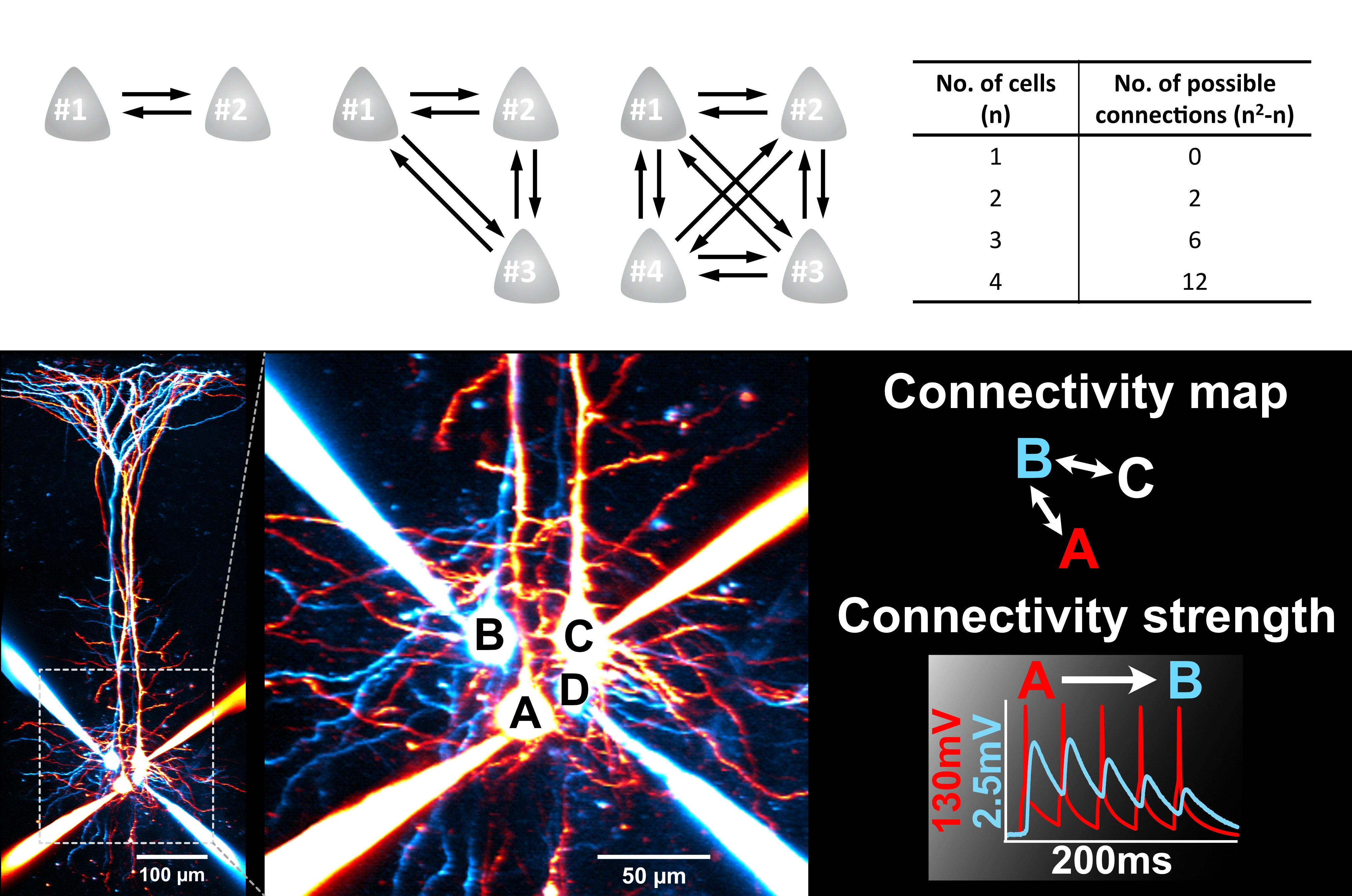
Whole-Cell Electrophysiology allows sub-millisecond and micro-volt electrical activities to be measured. These are crucial for understanding how neurons communicate via synapses and integrate inputs - neural encoding and information transfer.
Brain connections are sparse (~10% in the neocortex). Finding connections to examine synaptic transmission is therefore difficult. Being able to patch multiple cells enables us to find connections at a much faster rate, making the impossible experiments possible.
This state-of-art technique continuous to be the gold standard for mapping circuits.
Brain tissues are highly light scattering. They are difficult to image live without clearing methods used in fixed samples. 2PLSM tackles this problem by enabling ~0.5 µm imaging precision.
Compared to epifluorescence or confocal microscopy, 2PLSM uses two photons of lower energy (therefore light of higher wavelengths as E∝1/λ ) to excite the same fluorophores. The near-infrared light is scattered less and absorbed less, allowing it to penetrate deeper into tissues.
In addition, the needs for two photons for fluorophore excitation also means 2PLSM has an excellent built-in optical sectioning capability. This is vital for imaging micron-sized structures like the presynaptic terminals and dendritic spines, permitting molecular-cellular processes to be visualized at a fine scale.
To enable high throughput experiments, we make use of optogenetics. This involves expressing light-activated ion channels (opsin) in neurons. By shining the photoactivation laser at the cells expressing opsin (named ChETA on the right), action potential firing can be reliably and remotely triggered.
This method also allows synaptic transmission to be rapidly interrogated. By performing whole-cell patch of the postsynaptic cell and laser-activation of presynaptic cells, synaptic responses can be read out from the postsynaptic cell with electrophysiology granularity.
Unlocking the secrets of the brain is ambitious. Experimental designs are hence becoming increasingly sophisticated.
To cater for these experimental needs, we design our own light paths and custom-build with optical components. Our lab combines electrophysiology, 2PLSM and optogenetics, enabling neural communication recordings, imaging and laser-mediated manipulation to be performed simultaneously.
This also provides the means for us to develop new optical strategies tailored for neuroscience.
To understand the underlying fundament of brain cells and how their functions are mediated, we rely on molecular biology, live imaging and omics methods. These enable us to profile the molecular signatures that are dynamically regulated.
We are interested in all levels of molecular biology, including transcription, mRNA localization and translation and protein proteostasis. Through building these molecular atlases in health and disease conditions, we aim to develop novel gene therapy strategies to alleviate neurological disorders.
Drag & Drop Website Builder